MRI and Biopsy in Diagnosing Prostate Cancer
Role of MRI and Biopsy in Diagnosing Prostate Cancer
Magnetic Resonance Imaging (MRI) and biopsy are crucial tools in the diagnosis of prostate cancer, playing complementary roles in detecting and characterizing the disease. Prostate cancer is one of the most common cancers among men, and accurate diagnosis is essential for appropriate treatment planning.
MRI in Prostate Cancer Diagnosis:
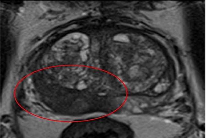
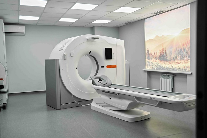
MRI has revolutionized the diagnosis and management of prostate cancer by providing detailed images of the prostate gland and surrounding tissues. Radiologists interpret these images to identify suspicious regions, which are often assigned a score based on the likelihood of cancer presence (PI-RADS score). MRI can help in:
- Detection: It can identify areas of suspicion within the prostate, guiding the decision for targeted biopsies.
- Localization: MRI assists in precisely locating suspicious regions, aiding in targeted biopsy planning.
- Risk Stratification: The PI-RADS scoring system helps assess the probability of malignancy, aiding in treatment decision-making.
- Staging: MRI can provide information about tumour extent and potential involvement of adjacent structures, guiding treatment strategies.
Biopsy in Prostate Cancer Diagnosis:
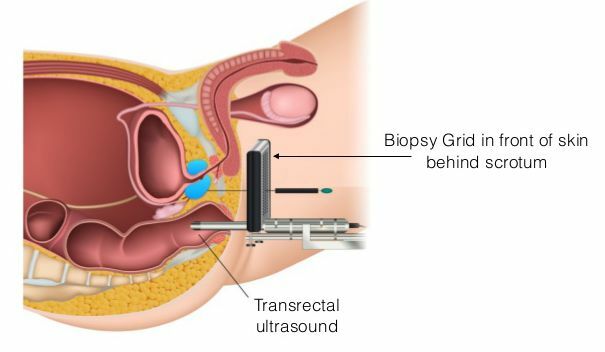
Prostate biopsy involves obtaining tissue samples from the prostate gland for histopathological analysis. This is usually done through a transperineal approach, often guided by MRI findings. While MRI provides valuable information, biopsy remains essential for definitive diagnosis. Biopsy is important for:
- Histopathological Confirmation: Tissue samples from biopsy allow pathologists to confirm the presence of cancer, assess its aggressiveness, and determine the Gleason score, which characterizes the tumour’s architecture.
- Sampling Accuracy: Biopsy provides direct tissue samples, allowing a detailed assessment of tumour characteristics, which might not be entirely captured by imaging alone.
- Treatment Planning: Biopsy results guide treatment decisions, including the choice between active surveillance, surgery, radiation therapy, or other interventions.
Combining MRI and Biopsy:
The integration of MRI and biopsy improves diagnostic accuracy. MRI-targeted biopsies, known as fusion or cognitive biopsies, use MRI images to guide the biopsy needle to suspicious areas identified on the MRI. This approach enhances the likelihood of detecting clinically significant tumours while minimizing the risk of missing aggressive cancers.
The synergy between MRI and biopsy significantly enhances the accuracy of diagnosing prostate cancer. MRI aids in identifying suspicious regions and guiding targeted biopsies, while biopsy provides the definitive histopathological confirmation necessary for treatment decisions. This combined approach leads to improved patient care by enabling tailored treatment strategies based on accurate cancer characterization.


